Over View
The instrument for quantitative cell biology at single-molecule detection.
Stimulated Emission Depletion (STED) is a powerful microscopy technique that allows for the observation of fluorescence structure with spatial resolution below the diffraction limit. The Alba-STED uses the pulsed excitation and pulsed depletion approach (pSTED) in combination with the digital frequency domain fluorescence lifetime imaging (FastFLIM) to record the time-resolved photons which allows for an increase in the image resolution and the separation of two labels with the same excitation wavelength.
Key Features of Alba-STED for FLIM/FFS:
- pSTED (Pulsed excitation and pulsed STED)
- FastFLIM for time-resolved pSTED acquisition
- Improved image resolution using the phasor plot
- Dual-label excitation
- Fast image acquisition (dwell time: 0.2 µs)
- High dynamic range (signal up to 60 million counts/s)
Measurements with Alba-STED for FLIM/FFS:
- 1p or 2p confocal images
- FLIM Fluorescence Lifetime Imaging (FastFLIM)
- PLIM, Phosphorescence Lifetime Imaging
- Polarization images
- Fluorescence Fluctuations Spectroscopy (FCS, FCCS, PCS, FLCS)
- RICS, N&B, Scanning FCS
- Burst Analysis
- FRET efficiency determination
Specifications for Alba-STED for FLIM/FFS
| Instrument Features | - Individual pinholes on each acquisition channel
- Computer-controlled selection of the pinhole variable aperture
- Computer-controlled positioning of the pinhole in the imaging plane
- Single-photon or multi-photon excitation
- Up to four channel data acquisition
- Auxiliary port for camera
|
| Acquisition and Analysis Software | |
| STED laser | Pulsed, 775 nmAverage output power: 1 W
Pulsewidth: about 600 ps
Repetition rate: 0-100 MHz; or Ext. CLK
Beam quality: M2 < 1.1, TEM00
Amplitude noise: < 4.0% rms |
| Excitation laser | Pulsed, 640 nmPulsewidth (at medium power): 40-90 ps
Repetition rate: 20, 50, 80 MHz; or Ext. CLK
Power (at 50 MHz): up to 5 mW |
| Laser Launcher | - Models for 3-, 4-, 6-laser. Light is delivered to the microscope through a single-mode fiber optic.
|
| FastFLIM | - 4 separate input signal
- Dynamic range: up to 15 million counts/s per channel
- Lifetime range: from picosecond to second
|
| Microscope | |
| Objectives | - Air objectives with 20X, 40X, 60X magnification and 1.5-8.1 working distances
- Oil immersion objectives, 1.4 NA and 60X (standard); other aperture available
- Water immersion objectives,1.2 NA 60X (standard), with coverslip correction (for 0.15-0.18 coverslip); other apertures available
|
| Stages | - Large distance movement (100x100x10 mm)
- Stepper motor-controlled XYZ stage
- Micro-distance movements
- XYZ piezo-controlled stage, 100x100x50 µm with 5 nm step resolution.
|
| Sample Holders | - Microwell plates
- Petri dishes
- Coverslip
|
| Light Detectors | - GaAs PMT (Hamamatsu H7422P models)
- Hybrid PMTs (Hamamatsu R10467U models)
- SPADs
|
| Image acquisition | FLIM acquisition: 0.2 µs dwell time |
| OS Requirements | |
| Power Requirements | - Universal power input: 110-240 V, 50/60 Hz, 400 VAC
|
| Dimensions | - 885 mm (L) x 600 mm (W) x 330 mm (H)
|
| Weight | |
Measurements for Alba-STED for FLIM/FFS
| FFS Measurements | - Fluorescence Correlation Spectroscopy (single- and cross-correlation)
- Photon Counting Histogram (PCH)
- FFS measurements at target XYZ locations in an image
- FLCS, Fluorescence Lifetime Correlation Spectroscopy
- Scanning FCS
- Number & Brightness (N&B)
- Raster Image Correlation Spectroscopy (RICS)
|
| Single-point Module Measurements | - Intensity
- Polarization
- Kinetics
- Lifetime
|
| Imaging Module Measurements (Single plan and z-stack) | - Intensity
- Polarization
- Ratiometric
- FLIM
|
| FLIM images (digital frequency-domain) (single plane and z-stack) | - Acquired in digital frequency-domain (DFD). The routine acquires simultaneously a FLIM image and a steady-state image.
|
| FLIM images time-domain (single plane and z-stack) | - Acquired in time-correlated single photon counting (TCSPC)
|
| Superresolution | |
| Single Molecule Module | - Burst Analysis
- FRET and Correlation Methods
- PIE-FRET Methods
|
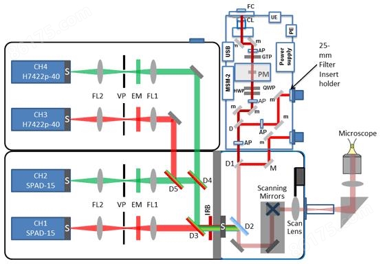
Schematic Diagram of Alba-STED for FLIM/FFS
Measurement Examples from Alba-STED for FLIM/FFS
Confocal (left) vs. pSTED (right) images of the actin labeled with the SiR dye in fixed glia cells, acquired by FastFLIM.
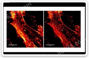
Confocal (left) vs. pSTED (right) images of the actin labeled with the SiR dye in fixed glia cells, acquired by FastFLIM.
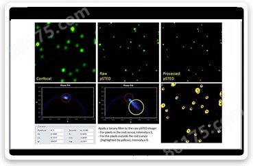
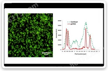
Confocal images of 60nm fluorescence beads (left); pSTED images (middle); sharpening the pSTED image using a binary filter based on the phasor plots (right).
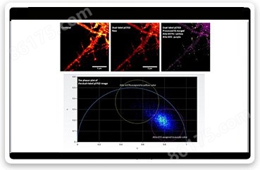
Dual labels can be separated using pSTED and FastFLIM. Atto 647N and Atto 655 were used as labels; they both are excited by the 640 nm laser. The two dyes are first separated using the phasor plots, and then assigned with two different false colors (Atto 647N - yellow, Atto 655 - purple) to produce the processed and merged pSTED image of the two labels.
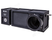
Options and Accessories available for Alba-STED for FLIM/FFS Non Descanned Detection (NDD) Port 
enlarge
Detection through the Non Descanned Detection Port (NDD) is used in conjunction with multiphoton excitation; the fluorescence photons generated in the excitation spot of the laser are scattered back and collected right after the objective (without passing through the optics in the scanning path).
The Figure displays the NDD port on the Nikon Model Ti microscope coupled to the Alba. A raiser is introduced on the Nikon microscope above the epifluorescence port for connecting the NDD port and adding the filters-cartridge where the dichroic filters for the NDD detection are inserted. The detectors are mounted on an orthogonal mount complete of dichroic and filter holders. The NDD port uses either GaAs PMTs or hybrid detectors. The output of the detectors is diverted either to the data acquisition unit.
Laser Scanning Transmitted Detection Module with a Photon Counting PMT
enlarge
T-PMT is a transmitted detection module with a photon counting PMT that works with inverted microscopes.
The module is installed beside the microscope transmission illumination lamp and features a manual slider for selecting between the PMT and the lamp illumination.
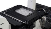
Microscope Stages
enlarge
XYZ-stepper motor controlled stage for Microwell plates (8-, 96- and 384-wellplate)
The XYZ stage provides high resolution, highly repeatable, and fast controls for the X, Y, and Z position of the microscope stage; it utilizes crossed-roller slides, a high-precision lead screw, and zero-backlash miniature geared DC servomotors for smooth and accurate motion. Controlled through the USB port, it is the ideal stage when measuring samples in a microwell plate.
VistaVision includes protocols for the automatic measurement at single points (FFS, lifetime, polarization); the user can select the sequential measurements on all the wells; alternatively, a set of wells can be selected for the measurements.